Home » Without Label » Blank Diagram Of A Long Bone / List 7-1 - Anatomy And Physiology with Ms. Dean at Camas ... / Skeletal system and long bone anatomy diagrams bundle.
Blank Diagram Of A Long Bone / List 7-1 - Anatomy And Physiology with Ms. Dean at Camas ... / Skeletal system and long bone anatomy diagrams bundle.
Blank Diagram Of A Long Bone / List 7-1 - Anatomy And Physiology with Ms. Dean at Camas ... / Skeletal system and long bone anatomy diagrams bundle.. Long, short, flat, irregular and sesamoid. It is placed laterally to tibia and is the most slender of all the long bones. Helps keep bones light in weight epiphyseal line line showing where growth plate used to be. Bones of the axial and appendicular skeleton. In long bones, as you move from the outer cortical compact bone to the inner medullary cavity, the bone transitions to spongy bone.
Shaft of a long bone. Smartdraw includes 1000s of professional healthcare and anatomy chart templates that you can modify and make your own. The femur and/or hip may fracture secondary to trauma, so understanding the femur bone anatomy is important. It is placed laterally to tibia and is the most slender of all the long bones. The tarsus or heel bone consist of 7 bones that make up the posterior part of the foot, that is present between the tibia, fibula and metatarsals.
Bone Trivia Questions - Bone Quizzes - Page 2 by ProProfs from www.proprofs.com The epiphyseal line is a remnant of an area that contained hyaline cartilage that grew. You need to get 100% to score the 10 points available. The end of the long bone is the epiphysis and the shaft is the diaphysis. It is 2 feet long and hollow, to make it lighter. Covers the surfaces of bones where they come together to form…. To skip right to the unlabeled diagram and quiz,. Short bones provide stability and support as well as. The bundle gives you access to a second set of diagrams, studying the anatomy of a long bone.
End of a long bone.
This is an online quiz called label the long bone. Helps keep bones light in weight epiphyseal line line showing where growth plate used to be. Blank bone diagram barca fontanacountryinn com. Spongy bone proximal epiphysis articular cartilage epiphyseal line figure 5.2a the structure of a long bone (humerus). In long bones, chondrocytes form a template of the hyaline cartilage diaphysis. The osteons are made up of the living osteocytes and mineral matrix which supplies blood. The calf bone or fibula is the smaller of the two bones that form the lower leg. Shaft of a long bone. Labeling diagrams and scientific illustrations is a valuable way to assess student learning, reinforce concepts, and integrate previous knowledge. Short bones provide stability and support as well as. Long, short, flat, irregular and sesamoid. Long bone diagram labeled find out more about long bone diagram labeled. The epiphyseal line is a remnant of an area that contained hyaline cartilage that grew.
Long bones function as rigid bars that move when muscles contract. This is a quiz called label the long bone and was created by member deanne1480 advertisement. Long, short, flat, irregular and sesamoid. The humerus is the long bone in the upper arm. Helps keep bones light in weight epiphyseal line line showing where growth plate used to be.
Long bone Parts Quiz from www.purposegames.com The anatomy of the femur can be divided into proximal, central, distal, and posterior parts. A long bone has two parts: Long bones include the humerus (upper arm), radius (forearm), ulna (forearm), femur (thigh), fibula (thin bone of the lower leg), tibia (shin bone) , phalanges (digital bones in the hands and feet), metacarpals (long bones within the hand), and metatarsals (long bones. Related posts of diagram of of a long bone bones and muscles anatomy. The diaphysis and the epiphysis. The epiphyseal line is a remnant of an area that contained hyaline cartilage that grew. The only short bones in the human skeleton are in the carpals of the wrists and the tarsals of the ankles. This is an online quiz called label the long bone.
The blood vessels inside a bone.
Bones of the axial and appendicular skeleton. Blank diagram of a long bone 6 3 bone structure anatomy. Long bone diagram labeled find out more about long bone diagram labeled. Covers the surfaces of bones where they come together to form…. These include the bones of the arms and legs. Helps keep bones light in weight epiphyseal line line showing where growth plate used to be. Long bones contain yellow bone marrow and red bone marrow, which produce blood cells. Long bones are one of the five bone types that are classified by shape. Short bones provide stability and support as well as. The ends of a long bone contain spongy bone and an epiphyseal line. The bundle gives you access to a second set of diagrams, studying the anatomy of a long bone. The end of the long bone is the epiphysis and the shaft is the diaphysis. The end of a long bone.
Long bones include the humerus (upper arm), radius (forearm), ulna (forearm), femur (thigh), fibula (thin bone of the lower leg), tibia (shin bone) , phalanges (digital bones in the hands and feet), metacarpals (long bones within the hand), and metatarsals (long bones. The structure of a long bone allows for the best visualization of all of the parts of a bone (figure 1). If you want a blank diagram to fill in manually, simply delete all of the data from the yellow boxes, then print your diagram. You need to get 100% to score the 10 points available. Long bones function as rigid bars that move when muscles contract.
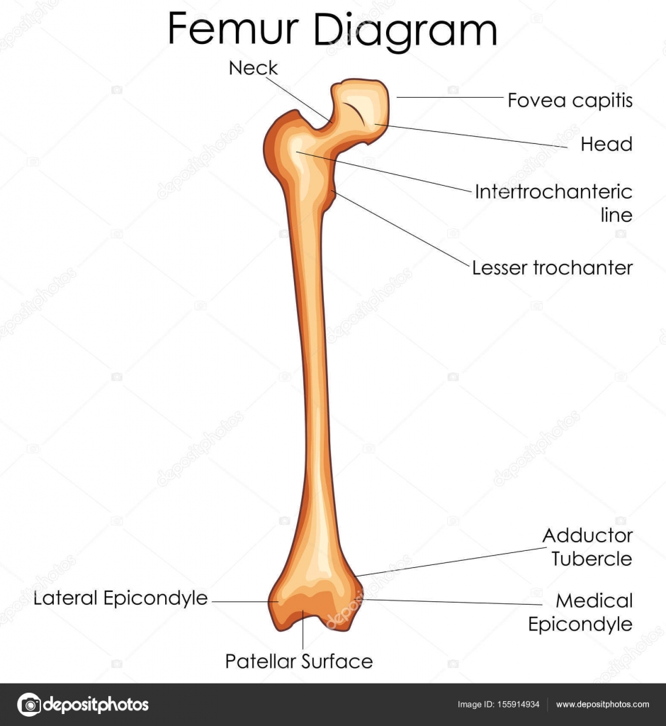
Femur bone diagram | Medical Education Chart of Biology ... from st3.depositphotos.com It is placed laterally to tibia and is the most slender of all the long bones. Related posts of long bone diagram labeled bone anatomy lecture. When a human finishes growing these parts fuse together. The common name of each bone is listed first, with the scientific name given in parenthesis. Short bones provide stability and support as well as. The only short bones in the human skeleton are in the carpals of the wrists and the tarsals of the ankles. Long bone diagram labeled find out more about long bone diagram labeled. Long bones are hard, dense bones that provide strength, structure, and mobility.
This is an online quiz called long bone diagram labeling.
Labeling diagrams and scientific illustrations is a valuable way to assess student learning, reinforce concepts, and integrate previous knowledge. Covers the surfaces of bones where they come together to form…. The femur is a type of long bone located in the thigh and is the largest bone of the skeletal system. It contains the bone marrow, one of the most important tissues in the vertebrate diagram of a typical long bone: Long, short, flat, irregular and sesamoid. The long bone diagram blank could be your desire when thinking of about bone. The end of a long bone. The largest bone in the body, the _____, is a long bone. There is a printable worksheet available for download here so you can take the quiz with pen and paper. Classification of bones from droualb.faculty.mjc.edu home » unlabelled » blank diagram of a long bone : Blank diagram of a long bone. Long bone shaft anatomy system human body anatomy diagram and. Blank bone diagram barca fontanacountryinn com.